Skin keratoma: causes, how dangerous is a neoplasm
The skin is the first and largest organ that faces the aggressive effects of environmental factors. Therefore, it is quite natural that specific protection mechanisms are provided in our body. One of them is the secretion of melanin, which protects the underlying layers of the dermis from the effects of ultraviolet radiation, and the constant renewal of the stratum corneum of the epidermis.

As a rule, such processes are clearly balanced, but under the influence of age-related changes in the mechanism of the flow of these reactions, failures occur, which often leads to various pathologies. These include keratoma of the skin.
Another name for this neoplasm is keratosis, in accordance with the international classification of the disease ICD, it was assigned the code D23. In the epidermis, cell renewal processes constantly occur. Replaced keratinocytes are replacing the dying and exfoliating structures of the stratum corneum.
Normally, these processes are balanced, but under the influence of a number of factors in the differentiation of keratinocytes fails, which leads to their enhanced division. As a result, a growth arises on the skin, resembling a wart , a birthmark or a birthmark protruding above the surface of the epidermis. This is how keratoma is formed.
Interesting fact
According to dermatologists, the main part of normal, room dust is made up of peeling skin particles.
According to experts, the starting factor for the onset of the disease is the influence of ultraviolet solar radiation. More precisely, the violation of the protective function of melanin. The fact is that with age, the level of its secretion, and accordingly the protective role it plays, is significantly reduced. It is these processes that are the main link in the pathogenesis of keratoma.
It is worth noting
Keratosis is referred to as benign neoplasms, since healthy cells take part in the process.
However, patients with a similar diagnosis do not exclude the likelihood of further malignancy, in other words, malignant degeneration of the cells of the epidermal cover. Therefore, patients with such an education on the body should be regularly examined by a dermatologist, therapist and oncologist.
For localization of keratoma there is no clearly defined specificity; theoretically, it can appear on any part of the body with the exception of mucous membranes, but mainly in the area exposed to sunlight. The color of the spot can also be different: from almost imperceptible at the initial stage to red or brown, sometimes with deep winding grooves on the surface.
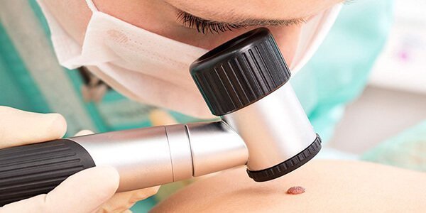
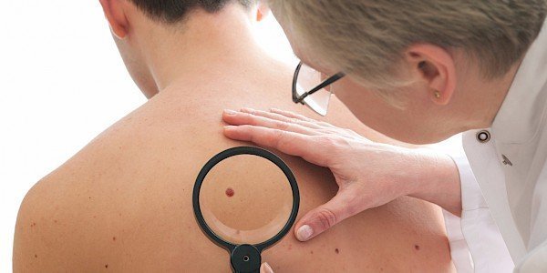
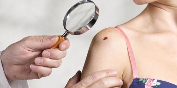
Photo keratoma is easy to find on the corresponding request on the network, but self-treatment is contraindicated. You should contact the appropriate specialist who will prescribe the necessary studies for accurate diagnosis and proper treatment (cryodestruction with liquid nitrogen, electrocoagulation, treatment of keratoma with laser or radio wave radiation, etc.).
Seborrheic keratoma and other types of the disease: description and distinguishing features
The classification of such neoplasms is based on clinical manifestations and the risk of degeneration into a malignant form. There are several types of keratomas: senile (or senile), follicular, seborrheic, horny, solar. Angiokeratome is also isolated in a separate group. Some forms can be combined, certain types are difficult to treat, others are prone to self-extinction.

Senile
This type of tumor is the most common, it is associated with age-related changes in the structure and blood flow in the epidermis. Pathology is characterized by the formation of protruding spots of various sizes (with a diameter of up to half a centimeter), mainly in open areas of the body (face, hands, chest, shoulders, etc.). Usually senile keratoma occurs in patients older than 40 - 50 years.
It is worth noting
With the late manifestation of this type of pathology (in patients after 50 years), the formation more often appears, on the contrary, on the skin, usually closed under clothing (abdomen, back, etc.).
Senile keratoma grows gradually, strongly rising above the surface of the skin, acquiring a grayish or brown tint. The consistency of the growth is soft, in some cases, when you press, there is pain or discomfort. This form of education leads to its frequent injury, which is accompanied by bleeding.
It is worth noting
Regular damage to senile keratoma increases the risk of malignant transformation.
Follicular
Occurs due to increased division of keratinocytes located in the area of hair follicles. Typically, the formation of color does not differ from the surrounding skin, its size rarely exceeds 15 mm, in the middle there is a small depression from which hair can grow. Characteristic places of localization - the face in the chin and scalp, much less often - the groin area, upper and lower limbs. The risk of malignancy is minimal, but this type of keratoma tends to recur frequently.
Seborrheic
In addition to impaired keratinocyte differentiation and melanogenesis, excessive sebum secretion lies in the pathogenesis of seborrheic keratoma. Accordingly, similar formations are localized in the area of concentration of the sebaceous glands (on the chest, back, scalp, etc.). In appearance, the outgrowth resembles a wart, differs in a yellowish tinge, the size usually exceeds 3 cm. The independent disappearance of keratoma is possible without any treatment, the probability of malignant transformation is almost minimal.

Horny
The name of this kind of pathology is fully consistent with the clinical picture. An outgrowth similar to a horn (the size can reach 10 cm or more), of a yellowish or grayish tint, sufficiently dense to the touch appears over the surface of the skin. The predominant sites of localization are the face (the region of the lips and lower eyelids), the area around the genitals.
It is worth noting
Compared to other types of the disease, horny keratoma is more prone to malignant degeneration.
Solar
Differs variable flow. Usually characterized by the appearance of pale red or pinkish spots covered with a scaly crust and surrounded by a slightly inflamed area of the epidermis. With equal probability, solar keratoma can spontaneously disappear, reappear after a long remission, or develop into a malignant neoplasm.
Angiokeratoma
In this case, the blood vessels of the subcutaneous tissue are involved in the pathological process, which affects the clinical picture of the disease. Occur single or multiple tumors, mainly in the region of the limbs, the body or external genital organs, ranging in size from 0.1 to 1 cm, sometimes growths can merge into one spot. The color of keratoma varies from red to dark brown or even black. Angiokeratoma requires mandatory treatment, but there is no risk of malignant malignancy.
At risk are all people older than 40 - 45 years, especially with a genetic predisposition to such skin diseases, pigmentation disorders. Also, keratoma appears more often in patients living in hot climates who have been under the open sun for a long time and do not use special sunscreens and lotions.
If seborrheic keratoma or another type of this disease appears on the skin, you should immediately consult a doctor.
A separate cause for concern is:
- a rapid increase in education, changing its shape and boundaries;
- spontaneous occurrence of other keratomas;
- blood or bleeding without previous damage;
- pain, itching, swelling and hyperemia in the area of appearance of keratoma.
To determine the form of keratoma, only a single examination and anamnesis is not enough. A dermatologist makes a sample of the material for biopsy. This examination allows to assess the risk of further adverse course of pathology. The determination of the method of treatment depends on the results of the tests.
Removal of keratomas: the main methods of treatment and prevention
Keratomas are not amenable to conservative therapy, some drugs and procedures are prescribed in addition when a malignant course of the disease is detected. But in most cases, laboratory tests indicate that there is no risk to the health and life of the patient.

Keratomas are removed by one of the following methods:
- Exposure to laser irradiation. Under the influence of ultrahigh temperatures, the neoplasm cells, roughly speaking, evaporate, which guarantees the absence of scars and the complete removal of growth. At the same time, the blood vessels coagulate, so the manipulation is not accompanied by bleeding, pain, and the recovery period does not exceed 7 - 10 days.
- Cryodestruction The destruction of keratoma occurs as a result of exposure to very low temperatures. For the manipulation of applied applications of liquid nitrogen. The procedure itself is painless, but after it is possible slight discomfort, redness of the skin. The overall healing process takes no more than 3-4 weeks.
- Use of a radio wave knife. Manipulation resembles the usual surgical removal of keratoma, but instead of a metal blade, the doctor uses a radio wave knife. This technique is characterized by rapid recovery, the absence of scars and scars, the minimum probability of secondary complications.
- Electrocoagulation. To remove keratomas apply electric current of a certain frequency. The procedure resembles cryodestruction, but current discharges are used instead of freezing. Regeneration of the skin in the treatment area takes up to 4 weeks.
It is worth noting
If a malignant tumor is suspected, keratomas are removed in a specialized clinic only surgically. In this way, the doctor can completely eliminate all reborn cells.
Some patients prefer treatment with traditional methods. The following recipes are quite effective:
- every day before bedtime it is necessary to lubricate keratomas with pure (or 1: 1 mixture with lemon) freshly squeezed parsley juice, by the way, this method is also suitable for bleaching freckles;
- in 1 liter of milk for 5-10 minutes, boil whole or chopped parsley root, leave overnight, strain and drink during the day;
- chop celandine herb into mush and mix with pork fat or petroleum jelly, apply as often as possible to keratoma;
- grind the aloe leaf, attach it to the neoplasm and leave it overnight, then remove the rest of the mass with the help of salicylic alcohol;
- mix baking soda and castor oil in a 1: 1 combination and apply regularly on keratome.
The main way to prevent such a disease is to protect the skin from the aggressive effects of ultraviolet radiation with creams, light clothing, etc. But the emergence of such education should not be a cause for panic. You should make an appointment with a doctor as soon as possible, undergo necessary examinations and carry out the removal of keratomas correctly, without the risk of complications and recurrences.