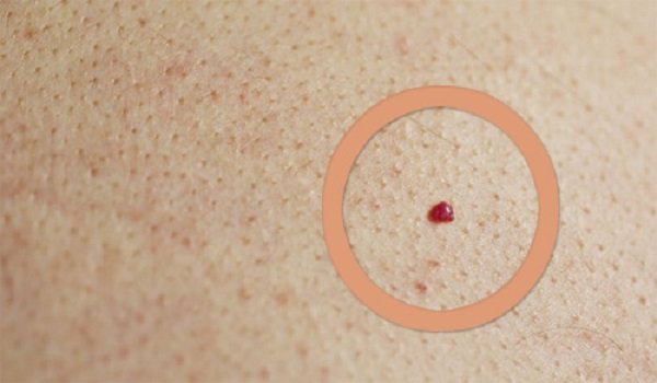
Red moles: causes, classification, clinical manifestations
Angiomas (this is how red moles are called in official medicine) are formed as a benign tumor of vascular origin. The basis of the pathogenesis of the disease is hyperplasia of blood vessels located in the subcutaneous tissue. The exact reasons for such changes are not fully known today, but most doctors associate them with systemic pathologies.

Angiomas are prone to benign course and do not pose a danger to human health. The risk of malignancy of tissues and malignant development increases only with a combination of several provoking factors (hereditary predisposition, aggressive exposure to ultraviolet radiation, etc.).
Such education can be both innate and acquired. According to the doctors, fetal hypoxia (even short-term) can cause angioma during intrauterine development. With oxygen starvation, the growing body seeks to fill the created deficit, which leads to a pathological proliferation of the circulatory network.
In some cases, the restoration of tissue respiration processes all returns to normal, sometimes, the angioma remains after the birth of the child. But this explains the spontaneous self-resolution of the disease and the disappearance of red spots or spots from the body of children as they grow up.
In adulthood, angio is associated with:
- failure of hormonal background during pregnancy, menopausal disorders, diseases of the endocrine system, taking oral contraceptive pills and other drugs containing hormones;
- autoimmune processes, when the circulatory network gets under the “sight” of a person’s own immune cells;
- pathologies of the digestive, urinary system;
- deficiency of ascorbic acid and other physiological angioprotectors;
- atherosclerotic changes, lipid metabolism disorders;
- genetic predisposition;
- mechanical damage to blood vessels due to injury;
- violations of the activity of the blood coagulation system.
It is worth noting
A severe viral or bacterial infection, prolonged exposure to the sun, contact allergies, exposure to the skin of toxins and other chemical irritants can provoke the growth of angiomas.
Depending on the nature of the neoplasm, angiomas are divided into hemangiomas, which are based on the proliferation of blood vessels, and lymphangiomas provoked by the pathology of the lymphatic network. The latter species occurs in isolated cases.
In form and size, these formations are classified into:
- the knobbly , angioma rises abruptly above the epidermal cover, is highly visible, usually has delineated rounded edges, and sometimes may resemble a hanging papilloma of intense color;
- nodular , small in size angioma, in size does not exceed a point or "asterisk";
- arachnid , has a flattened shape, visible network of blood vessels surrounding such a neoplasm;
- flat in appearance resembles a mole slightly elevated above the surface of the skin.
Depending on the number of blood vessels involved in the pathological process, angiomas can be:
- capillary , there is no specific localization, such formations are usually small in size, can be located anywhere on the body;
- cavernous , formed by rather large blood vessels, therefore it is formed mainly in areas richly supplied with blood (face, scalp).
It is worth noting
In some cases, cavernous angiomas may be located on the walls of internal organs.
- branchy , are a convex plexus of several formations;
- dotted , also called spider veins, look like the smallest points that do not rise above the surface of the skin.
In appearance, angiomas are similar to moles of bright red, brown or pink hue. They can appear at any age, even in infants. A distinctive feature is the blanching of the color with the pressure of a finger, then the color of the formation becomes the same.
Red moles on the body: localization features, treatment methods, use of alternative medicine
Typical sites of localization of angiomas are the face , including the eye area, the scalp. Less commonly, they occur on the chest, abdomen, and genital lips. In some cases, such formations extend to the subcutaneous tissue, muscle and connective tissue layer.

It is worth noting
In the vast majority of patients, red moles on the body are formed in the upper part of the body. Such features of the location are associated with the structure of the circulatory system and the intensity of blood flow.
Sometimes angiomas occur in early or adolescence, more often in adult women, since women are more susceptible to fluctuations in hormonal levels.
Today, medicine offers several ways to remove angiomas, which allow to remove the formation without unpleasant sensations, the subsequent formation of scars and scars and with minimal risk of postoperative complications.
The patient is asked to perform one of the following procedures:
- Laser removal . It is the safest and most modern method of getting rid of angiomas. The manipulation is performed under local anesthesia, but the whole procedure takes no longer than several minutes. Under the influence of laser radiation, the angioma completely “evaporates” without releasing blood and damaging the circulatory network.
- Moxibustion by radiowave, ultrasound and infrared therapy. The principle of such techniques has much in common with laser removal of angiomas . However, instead of the laser used other types of radiation. Such methods are quite effective, also painless, but the healing period of the skin takes longer.
- Cryodestruction using liquid nitrogen . Previously, this procedure was carried out quite often, but with the advent of other methods of hardware removal of the angioma, “freezing” is hardly used because of the long recovery period.
- Surgical removal of angiomas. This is almost a full-fledged operation, which is performed under local anesthesia, but only in a hospital. Such a procedure is indicated if the formation is dangerously malignant, born in an inaccessible place, or is too large.
From the popular methods of treatment, it is suggested to lubricate the angiomas with honey, onion juice, apply a slurry of grated radish or potatoes. It is also useful to take castor oil. They contribute to the strengthening of the vascular wall and the gradual disappearance of the angioma.
It is worth noting
Angiomas are absolutely benign education, but nevertheless you do not need to make a diagnosis on a photo from the Internet. Before removal it is necessary to consult a doctor.
Red moles on the skin: how dangerous education, especially lifestyle
If an angioma occurs in a newborn baby, doctors recommend refraining from any medical procedures. For most children, such educations dissolve independently, without affecting the functioning of internal organs and systems.

However, in both early and late age, red moles on the skin require removal in such cases:
- with a pronounced aesthetic defect due to the characteristics of localization;
- with permanent accidental damage to the formation;
- at risk of malignant rebirth.
In general, angiomas are not accompanied by the formation of secondary tumors, however, it is necessary to urgently sign up for a consultation with a doctor when the following symptoms appear:
- an increase in the size of angiomas;
- color change education;
- persistent itching;
- discharge of blood from the surface of the angioma.
Some patients experience recurrent itching in the area of vascular formation. This is usually associated with changes in hormonal levels. But often these symptoms are provoked by the further growth of the angioma and the involvement of other vessels in the pathological process. Therefore, it is better to consult a doctor and remove education.
By and large, red moles on the skin do not affect the lifestyle (of course, except when they are permanently injured). You can play sports, there are no strict restrictions in terms of nutrition, although it is better to adhere to the principles of proper diet.
However, ultraviolet is a very aggressive factor. In the area of hemangioma, certain violations occur in the secretion of the UV filter of melanin, therefore, when tanning, you need to use special protective creams and observe the time spent in the open sun.