Xanthelasma: etiology, clinical signs, diagnostic methods
It is no secret that various diseases of internal organs and vessels are reflected on the condition of the human skin. A striking example of such a dermatological pathology is xanthelasma (in some sources it is called flat xanthoma), one of the manifestations of systemic xanthomatosis.
This neoplasm has a certain localization, is not prone to malignant degeneration, but does not have a tendency to self-resolution and disappearance. To remove such a defect of the skin using methods of hardware and surgical cosmetology.
Although according to experts, a complete examination of the patient should be conducted to identify the most likely cause of xanthelasma. Treatment aimed at eliminating the etiological factor, almost 100% guarantees no relapse. Today, physicians are not completely sure what is the starting factor for the appearance of a similar formation on the skin. However, most doctors tend to consider xanthelasma a consequence of lipid metabolism disorders.
Histological examination of the biopsy specimen of such epidermal plaques clearly shows specific cells (later they were called xantom cells) in the form of phagocytes with one or several nuclei, foamy cytoplasm, and inclusions of different volume.
Typically, abnormalities in lipid metabolism are accompanied by the following systemic diseases:
- diabetes mellitus, usually in severe form or against the background of the lack of adequate insulin replacement therapy;
- various disorders of the liver - cirrhosis, hepatitis, etc .;
- pathologies affecting the functioning of the pancreas, for example, pancreatitis;
- thyroid disease, accompanied by a disorder of its endocrine function;
- overweight due to both nutritional errors and caused by pathological factors;
- genetically determined disorders of lipid metabolism.
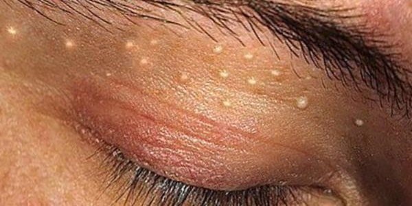
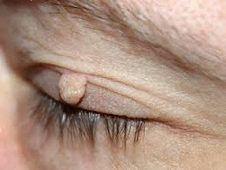
Xanthelasma is a flat plaque of yellowish or intense yellow color (in color it is brighter on the brighter side than the rest of the skin). Usually it is localized on the upper eyelid, closer to the inner corner of the eye. Although in some, quite rare cases, a similar formation occurs in the region of the lower eyelid.
The borders of xanthelasma are uneven, but clearly defined, it protrudes above the skin, the surface is smooth or covered with shallow folds. The size varies from 3-5 mm to 5 cm and more, in some patients the formation slowly but steadily grows, sometimes several plaques may merge together, the consistency is soft, pain is not noted when pressed. More often similar formations occur in women of middle and old age.
It is worth noting
In nearly half of patients with xanthelasma, lipid profile values are within the normal range. Therefore, this disease is more associated with various pathologies of the liver.
Education in the eye is a symptom of systemic xanthomatosis.
Other manifestations of this disease are:
- eruptive xanthomas - reddish papules, up to 1 cm in diameter, localized mainly on the extensor surfaces of the limbs, elbow, ankle, knee joints;
- tuberose xanthomas are dense spherical formations of a light brown color, usually located in the area of the buttocks, less often on the knees and elbows;
- tendon xanthomas form under the skin, often in the zone of the Achilles tendon;
- disseminated xanthomas, usually appear in patients with diabetes mellitus, are multiple eruptions up to 10 mm in size, yellow-brown in color, localized on sensitive skin (for example, in the armpits);
- juvenile xanthomas, occur in adolescence or in newborns, the formation of dense, spherical shape, located on the scalp, the region of the upper half of the body;
- warty xanthomas usually appear on the mucous membranes of the oral cavity, genital organs.
Diagnosis is carried out on the basis of data of external examination, anamnesis and histological examination. Microscopic examination of the biopsy shows accumulations of foam cells (sometimes they are arranged as cords), histocytes and lymphocytes, and also draw attention to the abundant capillary network.
Patients are required to prescribe lipid profile, laboratory and instrumental examination of the liver. Differential diagnosis of xanthelasma is carried out with benign skin syringoma, which has a similar clinical picture (the biopsy data differ) and sebaceous gland hyperplasia.
Laser Xanthelasma Removal and Other Treatment Methods

To date, there are several ways to completely get rid of xanthelasma with minimal risk to the patient. However, regardless of the method chosen, one should initially determine the treatment regimen for the underlying disease, which was the reason for the formation of xanthoma plaques in the eyelid area.
After the examination, determination of the glucose level, lipid profile parameters and liver function, appropriate medical therapy is prescribed. Otherwise, the likelihood of re-occurrence of tumors.
To get rid of xanthomas, you can use one of the following methods:
- Destruction of plaque cells with liquid nitrogen. This method is characterized by relatively low cost, however, due to the high morbidity of sensitive soft eyelid skin, the method is rarely used. Removal of xanthomas is carried out using an applicator dipped in liquid nitrogen. Due to the effects of low temperatures, necrosis and destruction of neoplasm cells occurs. After a few weeks, the structure of the skin is restored, and in place of the xanthoma there are no noticeable scars and scars.
- Radio wave exposure. A stream of radio waves is directed to the neoplasm, the parameters of which are chosen in such a way as to completely remove the plaque and not affect the healthy tissues located under and around it. The advantages of the method include the speed, painlessness, safety, no complications and side effects.
- Electrocoagulation. This technique of removing xanthoma can be accompanied by unpleasant sensations. Neoplasm cells are necrotized under the influence of electrical impulses.
- Surgery. It is shown in cases where the size or the presence of contraindications do not allow the tumor to be removed in another, safer way. Usually performed under local anesthesia, but accompanied by the risk of attaching a secondary bacterial infection. In addition, a significant drawback of surgical intervention is a long recovery period.
Laser removal of xanthelasma is currently considered the most modern and safe. This treatment method has several advantages. First of all, this is an impact only on abnormal cells without affecting the nearby healthy ones. This is especially important given the localization features of such a neoplasm.
Also, laser exposure is almost painless, does not cause bleeding due to simultaneous coagulation of blood vessels. The procedure creates high temperatures, which eliminates the likelihood of bacterial contamination. Wound healing after the procedure takes a couple of days, and the full process of skin recovery lasts up to several weeks.
However, the removal of xanthelasma laser has a significant drawback. Exposure to direct sunlight should be avoided for a certain period of time (usually 10–14 days). Therefore, it is better to postpone the procedure in the fall, winter, or use protective glasses.
It is worth noting
Methods of hardware cosmetology are effective for xanthelasma of relatively small size. If the plaques are fused with each other, doctors recommend resorting to surgery.
Removal of xanthelasma by laser, electrocoagulation, radio wave exposure is not recommended during pregnancy, lactation, patients with a tendency to bleeding implanted by pacemakers.
The procedures are carried out with caution in case of various dermatological diseases in an active form. Often, to eliminate possible pain and discomfort, anesthetic is injected before manipulation, making sure that there is no allergy to the drug used.
Xanthelasma century: treatment with alternative medicine, prevention, the cost of hardware therapy

A simple flat xanthoma of the eyelids is perfectly safe and will not lead to the formation of a malignant neoplasm. However, such a plaque may indicate serious violations of fat metabolism. Folk healers recommend starting treatment with vascular and liver cleansing.
So, the following recipes will do:
- Mix freshly squeezed onion juice with honey in a 1: 1 ratio. Take daily a tablespoon three times a day or before meals, or 2-3 hours after.
- Head garlic pour a glass of olive oil and clean in a cool place. You can start the reception the next day by mixing garlic oil with lemon juice (pour a teaspoonful of each drink into a glass), drink it three times a day 30 minutes before meals.
- Mix chamomile, tutsan, immortelle and birch buds (about 100 g each), grind in a meat grinder and add a little honey. Every evening, pour 2 tablespoons. mix half a liter of boiling water, filter in the morning and drink in two doses on an empty stomach - morning and evening.
- To cleanse the liver and improve its performance, it is necessary to mix mummy and aloe juice bought at a pharmacy in a ratio of 1:30. Take a teaspoon before breakfast and dinner, the course of treatment is 2 weeks, then a break of 14 days.
- Grind the grated or grinder horseradish root, squeeze out the juice and mix with honey in the same ratio. Take a tablespoon three times a day 20 minutes before meals.
From external agents, it is recommended to prepare a mixture of egg white, a teaspoon of honey and flour (approximately 10-15 g to obtain a uniform dense consistency). Mix all the ingredients and form small cakes, put them on the tumor for 15-20 minutes. Perform the procedure daily until complete disappearance of xanthelasma. Prevention of the occurrence of such plaques in the eye area is to follow an appropriate diet.
It is necessary to exclude all foods with a high content of cholesterol, fatty foods, sweets, soda, etc. It should be remembered that improper nutrition and overweight are the first steps to atherosclerosis, and the regenerative abilities of the liver are not unlimited. They recommend replacing coffee, tea and other drinks with wild rose, consume more vegetables and fruits, instead of sugar - dried fruits. Also shown is physical activity. After 35-40 years old, you should be examined regularly by a doctor or have a blood test and a lipid profile.
Xanthelasma eyelid is quite noticeable cosmetic defect, so most women go to the doctor in the early stages of plaque formation. The cost of consultation and examination ranges from 500-1500 rubles, hardware removal will cost around 1100-2000 rubles, depending on the chosen method of treatment. This amount does not include the cost of drugs prescribed for the correction of lipid metabolism and other associated diseases.
